EEG Test: What It Is, How It Works and What to Expect
An EEG is a painless test that records the electrical activity of your brain. Small metal discs called electrodes sit on your scalp and pick up the tiny signals your brain cells send to each other. A machine turns those signals into wavy lines on a screen, and a specialist reads the pattern. Doctors use this test most often to look for seizures and epilepsy, but it also helps with sleep problems, confusion and some other brain conditions. The Cleveland Clinic describes it as a safe test that can be done while you are awake or asleep.
In this article:
- What Is an EEG?
- How Does an EEG Work?
- Why Is an EEG Done?
- Types of EEG Tests
- What Happens During an EEG?
- How to Prepare for an EEG
- Brain Waves: What the Test Measures
- EEG vs CT Scan vs MRI
- Risks and Side Effects
- Understanding Your EEG Results
- Frequently Asked Questions
What Is an EEG?
An EEG, short for electroencephalogram, is a test that measures the electrical activity in your brain. Your brain cells, called neurons, communicate through tiny electrical impulses. The test records those impulses and shows them as brain waves.
Think of it as an ECG for the brain. An ECG records the electrical rhythm of your heart, while this test records the electrical rhythm of your brain. It shows how the brain is working from moment to moment, not what it looks like inside.
The test is non-invasive, which means nothing goes into your body and no electricity is sent into your head. The electrodes only listen to the signals your brain already produces. That is why the test is safe for people of all ages, from babies to older adults, and why it can be repeated when doctors need to track changes over time.
How Does an EEG Work?
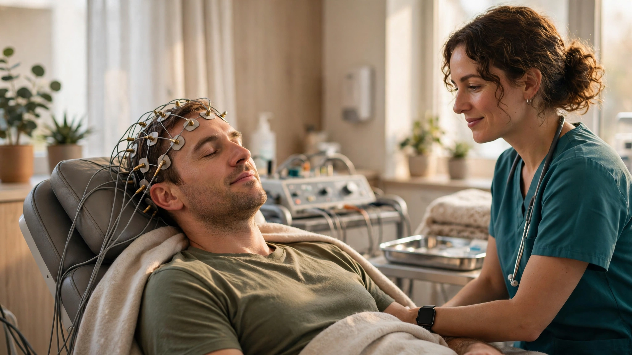
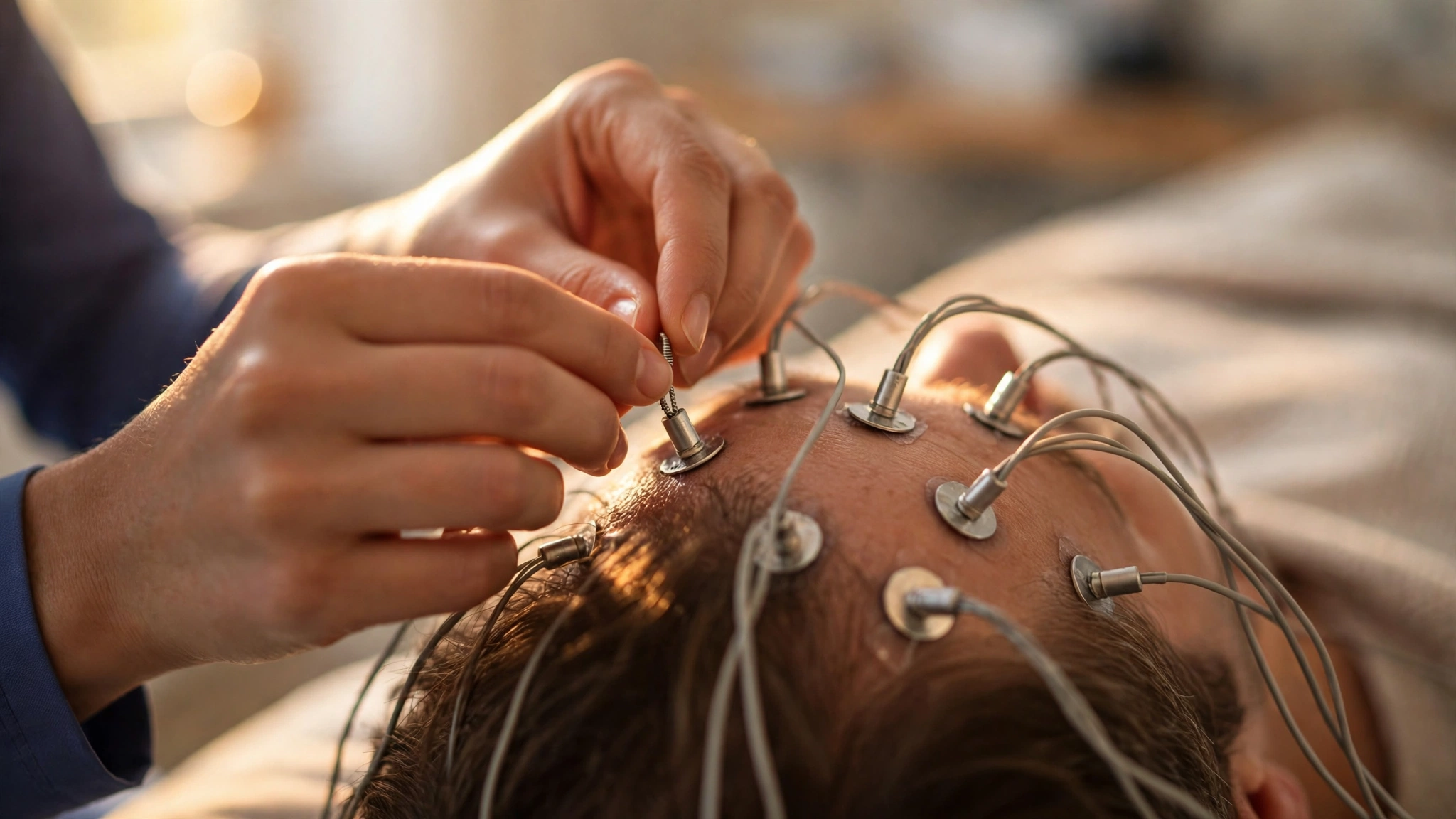
The test works in a simple chain of steps. First, a technologist places about 20 small metal discs on your scalp, spaced in a standard pattern. A sticky paste or glue holds each disc in place, and thin wires connect them to a recording machine.
Next, the discs detect the tiny electrical charges from your brain cells. These signals are far too small to feel, so you will not notice them at all. The machine amplifies the signals millions of times and draws them as wavy lines on a screen or on paper.
Then, the pattern changes as your brain state changes. When you are awake and alert, the waves look different from when you are drowsy or asleep. A neurologist studies these patterns, looking for waves that are too slow, too fast, or shaped in ways that suggest a problem.
Why Is an EEG Done?
Doctors order this test for several reasons, and seizures are the most common one. If you have had a seizure, a blackout, or a strange episode that might have been a seizure, the test can show whether abnormal electrical activity was involved. It also helps identify the type of epilepsy, which guides the choice of treatment.
- Unexplained confusion or memory loss. Sudden changes in thinking can come from abnormal brain activity, and the recording may show clues.
- Sleep disorders. The test can record brain waves during sleep, which helps diagnose conditions such as narcolepsy and some sleep-related seizures.
- Head injury or brain infection. After trauma or an infection such as encephalitis, doctors may check whether brain activity has changed.
- Dementia and other brain diseases. Some conditions change the speed and shape of brain waves in recognizable ways.
- Monitoring during surgery or in a coma. The test can track brain activity while a patient is under anesthesia or in a coma.
Doctors may also repeat the test to see whether a treatment is working.
Types of EEG Tests
Not every test of this kind looks the same. Your doctor picks the type that matches the question they need to answer. The main types are:
Routine EEG
This is the most common type. It takes place in a clinic or hospital and lasts about 20 to 40 minutes. You rest quietly with your eyes open or closed while the machine records. The technologist may ask you to breathe deeply for a few minutes or look at a flashing light, because these actions can bring out patterns that do not show up at rest.
Sleep-Deprived EEG
For this version, you stay awake for part or all of the night before the test. Being tired makes abnormal activity more likely to appear, which helps doctors catch seizures that a routine recording might miss. You may be asked to fall asleep during the test itself.
Ambulatory EEG
This version records for a longer period, often 24 to 72 hours, while you go about your normal day. You wear a small recorder on a belt or in a bag, with wires running to the electrodes under a cap or bandage. You keep a diary of what you do and feel, so doctors can match any symptoms to the recording.
Video EEG
This type pairs the recording with continuous video in a hospital room. It lasts from several hours to several days. Doctors use it when they need to see exactly what happens during an episode and where in the brain it starts. It is often part of the work-up before epilepsy surgery. Mayo Clinic explains that this longer monitoring helps pinpoint the source of seizures.
Prolonged or Continuous Monitoring
In intensive care, the recording may run for days to watch for silent seizures in patients who cannot report symptoms. This is common after a serious brain injury or during a coma.
What Happens During an EEG?
A routine test is simple and calm. Here is what usually happens, step by step:
- You settle in. You sit in a reclining chair or lie on a bed in a quiet, dimly lit room.
- Electrodes go on. The technologist measures your head and marks spots with a washable pencil. Each spot is cleaned, then a small disc is attached with paste or glue. About 20 discs are used, and fitting them takes 10 to 20 minutes.
- The recording starts. You rest with your eyes closed and stay as still as you can. Blinking, chewing and moving can blur the recording, so the technologist may remind you to relax.
- Simple tasks follow. You may be asked to open and close your eyes, breathe deeply and quickly for about three minutes, or watch a light that flashes at different speeds.
- The electrodes come off. The paste is washed out with warm water. The whole visit usually takes 60 to 90 minutes, including setup time.
How to Prepare for an EEG
Most people need little preparation, but a few steps help the test go smoothly:
- Wash your hair the night before. Clean hair helps the electrodes stick. Do not use conditioner, gel, spray or other styling products, because they can block the signal.
- Tell your doctor about your medicines. Some medicines affect brain waves, so your doctor may ask you to skip a dose or take it as usual. Never stop a medicine on your own.
- Follow sleep instructions. If you are booked for a sleep-deprived test, your doctor will tell you how much sleep to skip. For a routine test, a normal night of sleep is fine.
If you have a seizure disorder, tell the technologist before the test starts. Deep breathing and flashing lights can trigger a seizure in some people with epilepsy, and the team will be ready to help if that happens.
Brain Waves: What the Test Measures
The recording shows several kinds of brain waves, each linked to a different state. The main ones are:
- Delta – Slowest, under 4 Hz – Deep sleep
- Theta – 4 to 8 Hz – Drowsiness and light sleep
- Alpha – 8 to 13 Hz – Relaxed, eyes closed, awake
- Beta – 13 to 30 Hz – Alert, thinking, focused
- Gamma – Over 30 Hz – Intense focus and memory tasks
In a healthy brain, these waves shift smoothly as you move between waking, relaxing and sleeping. Abnormal patterns include waves that are too slow for your age, sudden spikes, or sharp waves that appear in one area. Spikes and sharp waves often point to a seizure disorder, while slow waves across the whole brain can suggest drowsiness, medicine effects or a more general brain problem.
EEG vs CT Scan vs MRI
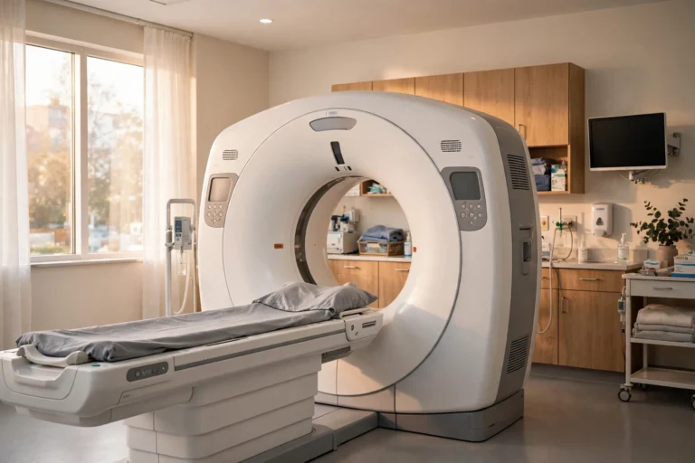
People often confuse this test with brain scans, but they measure different things. A CT scan or MRI shows the structure of the brain, such as a tumor, a bleed or a stroke. This test shows the function of the brain, meaning how its cells are firing right now.
- EEG – Electrical activity, brain waves – Seizures, epilepsy, sleep problems
- CT scan – Structure, fast images – Bleeding, fractures, urgent injury
- MRI – Detailed structure – Tumors, strokes, fine detail
Doctors often use them together. A scan may find a structural cause for seizures, while the recording shows whether seizures are still happening. Neither test replaces the other, and your doctor will explain which one answers your question.
The brain region that controls many of the rhythms this test records is the hypothalamus, which sets sleep cycles and body temperature. Our guide to nervous system regulation techniques also covers daily habits that support steady sleep and stress levels, and our tips on how to sleep better at night naturally can help if poor sleep is part of the picture.
Risks and Side Effects
This test is very safe, and most people feel nothing at all during it. A few minor effects are possible:
- Skin irritation. The paste or glue can leave the scalp slightly red or itchy where the discs sat. This fades quickly.
- Dizziness from deep breathing. Breathing fast for several minutes can make you feel light-headed. Tell the technologist if you feel unwell.
- A triggered seizure. In people with epilepsy, flashing lights or fast breathing can bring on a seizure. This is uncommon, and the team is trained to respond.
The test sends no electricity into your head and uses no radiation, so there is no long-term risk from the recording itself.
Understanding Your EEG Results
Results usually take a few days, because a neurologist must review the whole recording. The report describes whether the pattern was normal or abnormal, and what the abnormal parts might mean.
A normal result means the brain waves looked typical for your age and state during the recording. But a normal result does not rule out epilepsy. Seizure activity may simply not have happened during the test, which is why doctors sometimes order a longer recording. MedlinePlus notes this clearly: a normal test does not mean a seizure did not occur.
An abnormal result may show spikes, sharp waves or slow waves. The location and shape of these patterns help doctors narrow down the type of seizure disorder or the area involved. Abnormal does not always mean epilepsy, though. Sleep deprivation, medicines, low blood sugar and some infections can all change the pattern.
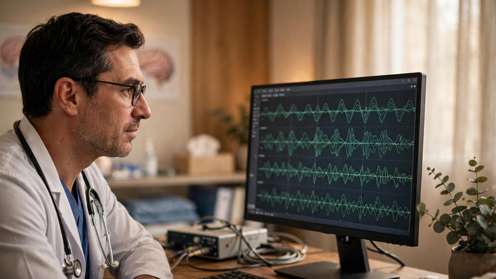
Your doctor will explain the result in the context of your symptoms and other tests. The recording is one piece of the picture, not a diagnosis on its own. If the result raises questions, the next step may be a longer recording, a scan, or a referral to a neurologist.
Frequently Asked Questions
What does an EEG test for?
This test checks the electrical activity of the brain. Doctors use it most often to diagnose epilepsy and other seizure disorders, and also to look into sleep problems, confusion, head injuries and brain infections.
Is an EEG painful?
No, the test is painless. The electrodes sit on the surface of your scalp and only record signals, so you feel nothing from them. The paste can feel cool and slightly sticky, and the glue may tug a little when it is removed, but there is no pain.
How long does an EEG take?
A routine test takes 20 to 40 minutes of recording, and the whole visit usually lasts 60 to 90 minutes with setup and cleanup. Longer versions last much more: an ambulatory test runs 24 to 72 hours at home, and video monitoring in hospital can last several days.
Do I need to shave my head for an EEG?
No, you do not need to shave your head. The electrodes attach to the scalp through your hair using paste or glue. Washing your hair the night before, without conditioner or styling products, is enough to help them stick.
Can an EEG detect a brain tumor?
This test cannot diagnose a tumor on its own. It may show abnormal activity near a growth, but a tumor is found with a scan such as a CT or MRI. Doctors use the two kinds of tests together when they suspect a structural problem.
What should I avoid before an EEG?
Wash your hair the night before and skip conditioner and styling products. Avoid caffeine on the day if your doctor advises it, and follow any sleep instructions you were given. Tell your doctor about all medicines you take, and never stop a medicine without their advice.
Conclusion
An EEG is a safe, painless way to listen to your brain’s electrical activity. It helps doctors find seizures, understand sleep problems and track how the brain is working over time. The test itself is simple: electrodes on your scalp, a quiet room, and a recording that a specialist reads. If your doctor has ordered one, the best preparation is clean hair, a good night of sleep unless told otherwise, and a list of your symptoms to share. And if the result is normal but your symptoms continue, talk to your doctor about the next step, because one normal recording does not always tell the whole story.
This article is for general information only and is not medical advice. If you have an ongoing health concern, talk to a healthcare professional.